
3.2 Physiology of Vision
Vision is a tricky matter. When we see a pizza, a feather, or a hammer, we are actually seeing light bounce off that object and into our eye. Light enters the eye through the pupil, a tiny opening behind the cornea. The pupil regulates the amount of light entering the eye by contracting (getting smaller) in bright light and dilating (getting larger) in dimmer light. Once past the pupil, light passes through the lens, which focuses an image on a thin layer of cells in the back of the eye, called the retina.
Because we have two eyes in separate locations, the image focused on each retina is from a slightly different angle (binocular disparity), providing us with our depth perception of 3D space (binocular vision).
It is in the retina that light is transduced, or converted into electrical signals, by specialized cells called photoreceptors. The retina contains two main kinds of photoreceptors: rods and cones.
Rods are primarily responsible for our ability to see in dim light conditions, such as during the night. Cones, on the other hand, provide us with the ability to see color and fine detail when the light is brighter. The highest concentration of cones is found in the fovea (the central region of focus), while rods dominate the periphery. The difference in distribution can explain why looking directly at a dim star in the sky makes it seem to disappear; there are not enough rods to process the dim light!
Color vision

Our cones allow us to see details in normal light conditions, as well as color. We have cones that respond preferentially, not exclusively, for red, green, and blue (Svaetichin, 1955). This trichromatic theory is not new; it dates back to the early 19th century (Young, 1802; Von Helmholtz, 1867). This theory, however, does not explain the odd effect that occurs when we look at a white wall after staring at a picture for around 30 seconds. Try this: stare at the image of the green and black Canadian flag for 30 seconds and then immediately look at a sheet of white paper or a wall. According to the trichromatic theory of color vision, you should see white when you do that. Is that what you experienced? As you can see, the trichromatic theory doesn’t explain the afterimage you just witnessed. This is where the opponent-process theory comes in (Hering, 1920). This theory states that our cones send information to retinal ganglion cells that respond to pairs of colors (red-green, blue-yellow, black-white). These specialized cells take information from the cones and compute the difference between the two colors—a process that explains why we cannot see reddish-green or bluish-yellow, as well as why we see afterimages. Color deficient vision can result from issues with the cones or retinal ganglion cells involved in color vision.
Vision and the brain
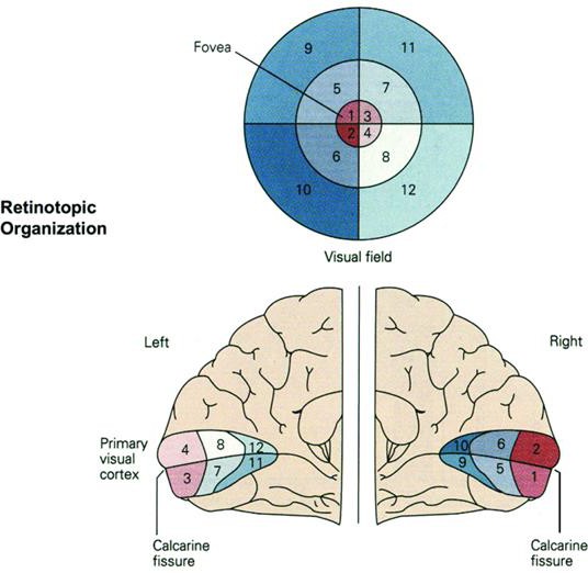
For visual information to reach the brain, the electrical signal is sent through a layer of cells in the retina, eventually traveling down the optic nerve. After passing through the thalamus, this signal makes it to the primary visual cortex, where initial information about light orientation and movement begin to come together (Hubel & Wiesel, 1962). The primary visual cortex is retinotopic: visual information falling on adjacent areas of the retina is processed in corresponding adjacent areas of the primary visual cortex (see the image below).
Information is then sent to various areas of the cortex for more complex processing. Some of these cortical regions are fairly specialized—for example:
- Fusiform Face Area (FFA): Detection (and possibly recognition) of faces.
- Extrastriate Body Area (EBA): Processing of non-facial body parts
- Parahippocampal Place Area (PPA): Conscious recognition of places
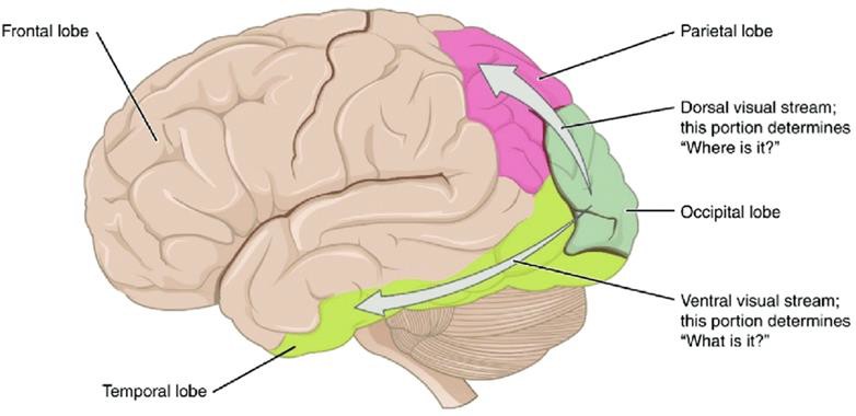
Damage to these areas of the cortex can potentially result in a specific kind of visual agnosia, whereby a person loses the ability to visually identify stimuli. A fitting example of this is illustrated in the writing of famous neurologist Dr. Oliver Sacks; he experienced prosopagnosia, the inability to recognize faces. These specialized regions for visual recognition comprise the ventral stream (also called the “what” pathway). Other areas involved in processing location and movement make up the dorsal stream (also called the “where” pathway). Together, these pathways process a large amount of information about visual stimuli (Goodale & Milner, 1992).
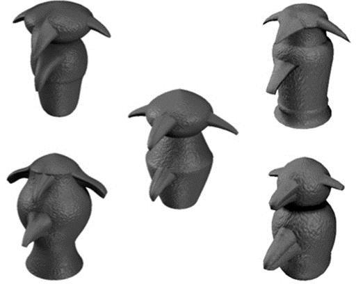
Of note is that there is some room for “experience-dependent plasticity” with these specialized regions; ;given experience, they can potentially be utilized to process other types of stimuli as well. In one study (Gauthier et al., 1999), researchers trained participants to differentiate unique objects with common features (sort of like faces) called “greebles” (see the image to the right). For participants trained extensively in this differentiation, they found enhanced activation in the fusiform face area compared to those without training.
Another study (Ison et al., 2015) measured individual neuron activity in the parahippocampal place area (usually dedicated to place recognition) and found increased activity for human faces that had been paired with specific locations but not other faces, suggesting a role in the formation of memories related to people in particular places.
Video: Visual stimuli and neural activity (the clicks in the video represent the action potentials of individual neurons) during the research of Ison et al. (2015).
Below, find a video from Crash Course Psychology explaining some of the concepts from this section regarding visual sensation and how it can be distinguished from our next topic, perception!
Cell layer in the back of the eye containing photoreceptors.
Our ability to perceive 3D and depth because of the difference between the images on each of our retinas.
A loss or impairment of the ability to recognize and understand visual stimuli.
A form of visual agnosia in which the ability to perceive and recognize faces is impaired.
Pathway of visual processing flowing from the occipital cortex to the temporal lobe, representing nameable visual features of objects. Also known as the “what” pathway.
Pathway of visual processing flowing from the primary visual cortex to the parietal lobe. Involved in processing object motion and location. Also known as the “where” pathway.
